Mini spinal cord grown in laboratory and healed
Scientists at Northwestern University created a mini model of the human spinal cord from stem cells, simulated traumatic injuries on it, and successfully tested a "dancing molecule" therapy that significantly reduced scarring and promoted n
A breakthrough in spinal cord injury research
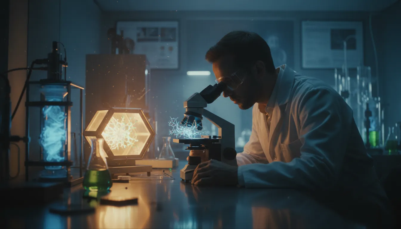
A team of scientists from Northwestern University in Illinois, USA, has achieved a significant milestone in biomedical research. They grew a three-millimetre organoid of the human spinal cord from embryonic stem cells — a miniature organ that closely mimics the structure and behaviour of the real spinal cord. They then simulated traumatic injuries on it and successfully tested an experimental treatment. The study was published on 11 February 2026 in the prestigious journal Nature Biomedical Engineering.
First organoid with immune cells
The research team, led by Professor Samuel I. Stupp and first author Nozomu Takata, cultured organoids from induced pluripotent stem cells for several months. During this period, neurons, astrocytes, and other cells typical of the spinal cord developed in them.
A key innovation was the first incorporation of microglia — immune cells of the central nervous system — into the spinal cord model. Microglia play a crucial role in the inflammatory response that follows spinal cord injury. Without them, the model could not accurately reproduce the actual course of trauma.
Simulation of real injuries
The scientists induced two types of injuries on the organoids that correspond to common clinical scenarios. Laceration — a cut injury with a scalpel mimicking surgical trauma — and contusion — a pressure injury simulating the consequences of a traffic accident or fall.
In both cases, the organoids accurately reproduced the key features of real spinal cord injury: cell death, inflammatory response and glial scar formation. This represents a dense mass of tissue that creates a physical and chemical barrier preventing nerve regeneration — and is one of the main reasons why paralysis after spinal cord injury is considered irreversible.
Dancing molecules yielded surprising results
The injured organoids were then treated with an experimental therapy called "dancing molecules". These are supramolecular peptides containing more than 100,000 molecules that, upon contact with tissue, form nanofibres that mimic the extracellular matrix of the spinal cord. Their rapid movement effectively activates cell receptors and stimulates regeneration.
The results were remarkable. The glial scar almost completely disappeared after treatment, and long projections — neurites — began to grow from the neurons, which are essential for restoring nerve connections. Nothing like this happened in untreated organoids.
"The dancing molecules sent long neurites from the surface of the organoid, but when we used molecules with less or no movement, we saw nothing," explained Professor Stupp.
The road to clinical trials
Dancing molecule therapy has previously proven effective in animal models — paralysed mice were able to walk again within four weeks after a single injection. The US Food and Drug Administration (FDA) has granted it orphan drug status, indicating that clinical trials in humans are imminent.
The organoid model opens up further possibilities. Scientists plan to create models of chronic spinal cord injuries and explore personalized medicine approaches using the patients' own stem cells. Such an approach could lead to tailored treatments without the need for animal testing — an important ethical and practical shift in medical research.