How Scientists Map the Brain's Wiring
Connectomics is the science of mapping every neural connection in a brain. From the 302-neuron worm mapped in the 1980s to the fruit fly's 130,000 neurons completed in 2024, researchers are building ever-larger wiring diagrams that could transform our understanding of neurological disease.
What Is a Connectome?
A connectome is a complete map of every neural connection inside a brain — a wiring diagram that shows which neurons talk to which, and through what kind of synapse. The term was coined independently in 2005 by neuroscientist Olaf Sporns at Indiana University and researcher Patric Hagmann at Lausanne University Hospital. The field built around this idea, connectomics, rests on a simple premise: to understand how a brain works, you first need to know how it is wired.
Think of it like trying to diagnose a fault in a city's electrical grid. Without a circuit diagram, every repair is guesswork. The same logic applies to the brain — and to the diseases that attack it.
From Worms to Flies: A Brief History
The first connectome took decades. In the 1970s, biologist Sydney Brenner and his team at the MRC Laboratory of Molecular Biology began slicing the roundworm Caenorhabditis elegans into ultra-thin sections, photographing each one under an electron microscope, and painstakingly tracing every neural pathway by hand. The result, published in 1986, mapped all 302 neurons and roughly 7,000 synapses in the worm's nervous system — the first complete wiring diagram of any animal.
It remained the only complete connectome for nearly four decades. Then in 2024, a large international consortium funded by the U.S. National Institutes of Health published the full connectome of the adult fruit fly (Drosophila melanogaster) — a staggering leap in complexity. The fly brain contains roughly 139,000 neurons and over 54 million synapses, published across nine papers in Nature.
How Brain Mapping Works
There is no single technique for mapping connectomes. The method depends on the scale researchers need.
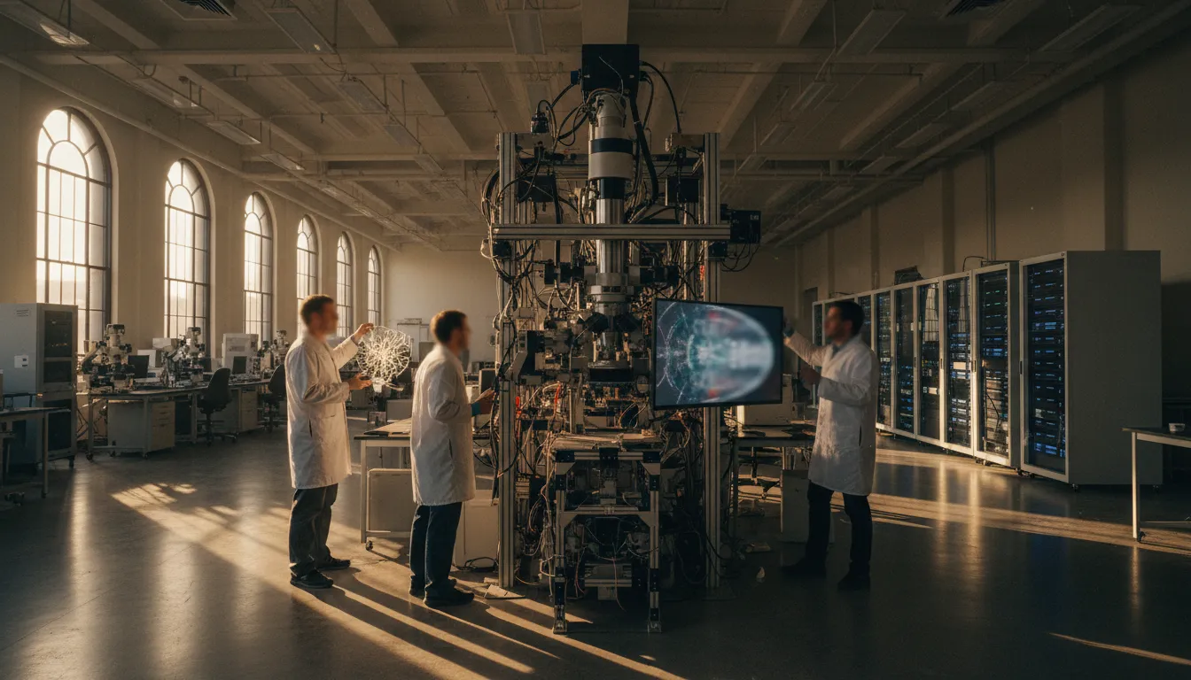
Electron Microscopy (Nano-Scale)
For neuron-by-neuron maps, scientists slice brain tissue into sections just tens of nanometers thick — far thinner than a human hair — and image each slice with an electron microscope. Software then stitches millions of images together, and researchers (often aided by AI) trace the path of every axon and dendrite through the stack. This is how both the worm and fly connectomes were built. It is extraordinarily detailed but slow and expensive.
Diffusion MRI (Macro-Scale)
For living human brains, researchers use diffusion-weighted magnetic resonance imaging (dMRI), which tracks the movement of water molecules along nerve fibers. Combined with tractography algorithms, dMRI can reconstruct the brain's major white-matter highways — the long-distance cables connecting distant regions. The Human Connectome Project, launched by the NIH in 2009 with $40 million in funding, used this approach to scan 1,200 healthy adults and identify 180 distinct cortical areas, including 97 previously unknown regions.
RNA Barcoding (The New Frontier)
A technique called Connectome-seq, published in Nature Methods in 2026, takes a radically different approach. Scientists assign each neuron a unique RNA "barcode." Engineered proteins carry these barcodes to the synapse, where pre-synaptic and post-synaptic tags sit side by side. Researchers then isolate synapses and use high-throughput sequencing to read which barcode pairs appear together — revealing direct connections between neurons without ever touching a microscope. In its first demonstration, the team mapped over 1,000 neurons in a mouse brain circuit and uncovered previously unknown connections between cell types.
Why It Matters
Connectome maps are not just academic exercises. By comparing the wiring of healthy brains with those affected by conditions such as Alzheimer's disease, schizophrenia, or autism, researchers hope to identify the specific circuit-level changes that drive these disorders. The fruit fly connectome, for example, already allows scientists to trace the exact neural pathways behind behaviors like navigation and decision-making — circuits with surprising parallels to mammalian brains.
The ultimate goal is a complete connectome of the human brain, with its roughly 86 billion neurons and 100 trillion synapses. At current speeds, that remains far off. But each new technique — from faster electron microscopy to RNA barcoding — shrinks the timeline. What once took Brenner's team a decade to map in a worm could eventually be accomplished across an entire human brain, transforming neurology from educated guesswork into precision engineering.